

3D X-ray diagnostics LiderDent
Diagnostic Imaging
X-ray diagnostics
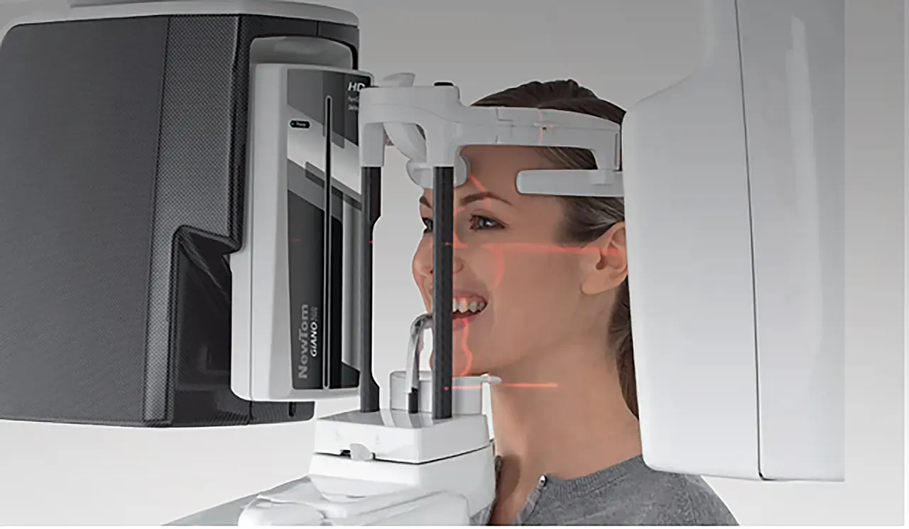
Polyclinic LiderDent uses the most modern device for X-ray diagnostics in order to provide its patients with all the necessary X-ray digital diagnostics with a minimum dose of radiation and high-quality reproduction of images. The device offers a wide range of 2D and 3D images of different imaging fields. Visualization and diagnostic plan is carried out in advanced dental software.
Panoramic X-ray or Orthopantomogram
OPG represents a panoramic 2D imaging of both the upper and lower jaws and surrounding bones. This image is mandatory during the patient’s first visit and is used to assess the teeth condition, bone structure, and periodontium (the supporting structure for the teeth). The imaging process is completely painless and comfortable for patients as both the sensor and the X-ray source are positioned outside the mouth, rotating around the patient's head.
Temporomandibular Joint (TMJ) Imaging
Targeted imaging of the bone structures of both temporomandibular joints with open and closed mouth is used in diagnosing temporomandibular joint diseases. It is available in both 2D or 3D.
Teleroentgenography
Teleroentgenography is a 2D image used in orthodontic diagnostics. It usually consists of an LL (latero-lateral) imaging and an AP (anteroposterior) imaging, especially in patients whose skeletal growth is being monitored. The lateral view provides information about the relationship between the upper and lower jaws in all spatial planes, is used to monitor a child's growth, and to assess the initiation of orthodontic therapy. The AP imaging is used in diagnosing facial asymmetry and preparing patients for orthognathic surgery. Additionally, it is possible to perform an X-ray of a person’s hand in the clinic, which is used in diagnosing the completion of growth and development. This image is meaningless without software analysis, through which the orthodontist makes a diagnosis and treatment plan.
CBCT - Cone Beam Computer Tomography
Cone beam is a 3D imaging method using cone beam computed tomography, where the image creates a reconstructed three-dimensional image of the scanned area, which greatly facilitates diagnostic work and therapy planning.
3D imaging has advanced significantly in the last two decades and has found application in orthodontics, oral and maxillofacial surgery, and especially in situations that require the understanding of complex anatomical relationships and the surrounding structures of the maxillofacial skeleton. The device allows us to scan various parts of the face. The key advantage of CBCT is its high-resolution images at relatively low radiation doses. This imaging is completely painless and void of discomfort for the patient.
Paranasal Sinus Imaging
3D imaging of the maxillary sinuses is used in diagnosing sinus disease in otorhinolaryngology. This imaging is completely painless and without discomfort for the patient.
Airway Analysis for Air Passage Patency
Using CBCT machine/scanner in a specific software program, narrowing of the airways can be visualized. This provides a static image of the airways, thereby identifying patients with a high predisposition to obstructive sleep apnoea. It is very important to analyse the airway before orthognathic therapy to precisely prepare and plan the therapy for the surgical movement of the lower jaw.
In the Liderdent Polyclinic, the X-ray device is located in the basement of the office. Immobile or immobile patients and patients in wheelchairs cannot access this device. We refer these patients to cooperating institutions.
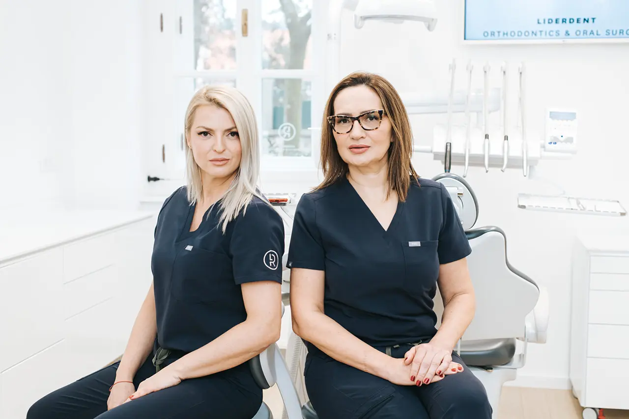
EXPERT WORD
COMPREHENSIVE PRistup
Our mission is to create small masterpieces and, with the method of 3D visualization and extensive diagnostic protocols that include a multidisciplinary approach, provide clients with excellent results of beauty, health and function .
– Renata Kevilj Gospić, PhD, MSc, DMD
– Lidija Jozinović, MD
HOW TO START?
The first step to a more beautiful smile
Consultations with a dentist are the first step towards a more beautiful smile – a smile that leaves a positive impression on other people, makes us more confident and full of confidence. Based on quality diagnostics, we will propose an individual therapy plan oriented towards achieving complete aesthetic and functional rehabilitation. In the safe hands of our doctors, we provide our clients with impeccable results with an individual approach, knowledge, and love for work.
THANK YOU TO THE KIND AND ALWAYS SMILING STAFF, AND ESPECIALLY TO DR. JOZINOVIĆ FOR BRINGING A SMILE BACK TO MY FACE!
SHE MADE AN EXCELLENT, INCONSPICUOUS IMPLANT, I CAN FINALLY SMILE WITHOUT HIDING!
Anthony T.
A big thank you to Dr. Renata for her exceptional professionalism and for everything she has done for me. Wonderful practice and wonderful staff! It was a pleasure to be your patient!
Ivan B.
service, professionalism, effort, commitment. Maximum effort was made to achieve excellent results. Definitely a recommendation to anyone who wants to achieve the best.
Vedran B.
I am very pleased with the service. very pleasant atmosphere and beautiful ambience. The doctor is very professional and nice. my warm recommendations.
Nancy M.
Thanks for the great change! I am very pleased and Believe that you will satisfy all your future clients.
Lucia B.
